
Page 40 - ITU KALEIDOSCOPE, ATLANTA 2019
P. 40
2019 ITU Kaleidoscope Academic Conference
Our ultimate goal is to create a visible lighting strategy with
different frequencies, waveforms and duration to prevent,
relieve and treat depression, mania, Alzheimer's disease,
preferably either in the hospital or nursing home.
Unlike mice, detecting human electroencephalogram(EEG)
signals relies on non-invasive devices, such as the open-
source brain-computer interface (OpenSCI) system [11].
Both commercial software and open-source software can be
used in the analysis of collected EEG signals for verification
in conjunction with life sciences and medical
professionals[12]. This software and the hardware provide
non-invasive methods to sample the electrical activities of
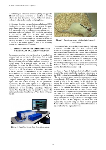
the body and brain of human beings. These methods are not Figure 5 - Experiment mouse with implanted electrode
as precise as the method applied to mice in the following in hippocampus
sections but conform to medical ethics.
Two groups of mice were used in the experiments. Following
3. DESCRIPTION OF THE EXPERIMENT AND a standard procedure, the mice were implanted with
PRELIMINARY ANALYSIS OF THE DATA electrodes in the hippocampus of the brain. After surgery, the
mice were allowed to recover for a week. After restoration,
The common practice is to use the animal to conduct the the OmniPlex Neural Data Acquisition system of Plexon was
research work and confirm the effectiveness first, to avoid used to collect local field potential signals for 15 minutes as
problems such as high uncertainty and inconsistency for reference. Then, the modulated 40Hz flashing LED lamp
direct application to human beings, and most importantly the was turned on to radiate the mice for 15 minutes, and the
ethical issues. As a concept study, we used 40 Hz local field potential signal was collected at the same time.
scintillation frequency for this preliminary experiment in Data was analyzed using NeuroExplorer, which is widely
visible light irradiation on mice, motivated by the work from used in the field of neuroscience [13].
Tsai and et. al [2]. The advantage of the mouse experiment
is that we can use the multichannel in-vivo recording to As shown in Figure 6, the brain electric local field potential
record and monitor the point activity of the neuron group signal of the mouse exhibited a significant enhancement in
directly inside the brain to obtain the local field potential the 20 Hz portion in the modulated visible light irradiation.
(LFP) signal of a certain brain region (hippocampus). The two subgraphs above show the comparison of power
Compared with the signals acquired outside the skull, it has spectral density (PSD) analysis results of local field potential
higher time and spatial accuracy. In our preliminary signals of mice in the two groups. The left side is the PSD
experiments, the field status signals of the hippocampus during the radiation and the right is the PSD before the
associated with learning and memory were collected. The radiation. It can be clearly seen that the hippocampal area of
multichannel in-vivo recording technology, OmniPlex mice in the radiation has obvious discharge and energy
Neural Data Acquisition, and experiments on mice with response at the frequency of 20Hz. The third subgraph shows
implanted electrodes for collecting LFP are shown in Figures the heatmap of the hippocampal region of mice with changes
4 and 5. over time. The left side is the heatmap during the radiation
and the right is the heatmap before the radiation. It can also
easily display that the energy distribution of the hippocampal
region of mice at the frequency of 20Hz has been
significantly enhanced during the whole radiation period.
Here, we have observed the results yet lots of work needs to
be done to offer the explanation why.
Figure 4 - OmniPlex Neural Data Acquisition system
– 20 –