
Page 104 - AI for Good Innovate for Impact
P. 104
AI for Good Innovate for Impact
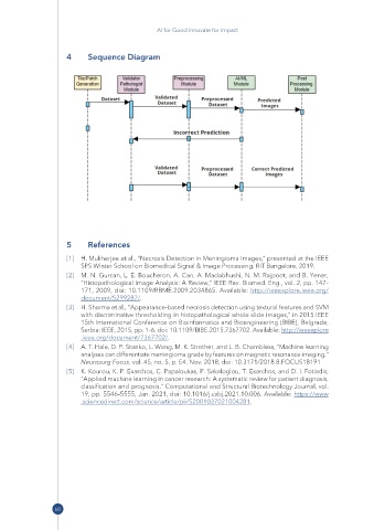
4 Sequence Diagram
5 References
[1] H. Mukherjee et al., “Necrosis Detection in Meningioma Images,” presented at the IEEE
SPS Winter School on Biomedical Signal & Image Processing, RIT Bangalore, 2019.
[2] M. N. Gurcan, L. E. Boucheron, A. Can, A. Madabhushi, N. M. Rajpoot, and B. Yener,
“Histopathological Image Analysis: A Review,” IEEE Rev. Biomed. Eng., vol. 2, pp. 147–
171, 2009, doi: 10.1109/RBME.2009.2034865. Available: http:// ieeexplore .ieee .org/
document/ 5299287/ .
[3] H. Sharma et al., “Appearance-based necrosis detection using textural features and SVM
with discriminative thresholding in histopathological whole slide images,” in 2015 IEEE
15th International Conference on Bioinformatics and Bioengineering (BIBE), Belgrade,
Serbia: IEEE, 2015, pp. 1–6. doi: 10.1109/BIBE.2015.7367702. Available: http:// ieeexplore
.ieee .org/ document/ 7367702/ .
[4] A. T. Hale, D. P. Stonko, L. Wang, M. K. Strother, and L. B. Chambless, “Machine learning
analyses can differentiate meningioma grade by features on magnetic resonance imaging,”
Neurosurg Focus, vol. 45, no. 5, p. E4, Nov. 2018, doi: 10.3171/2018.8.FOCUS18191
[5] K. Kourou, K. P. Exarchos, C. Papaloukas, P. Sakaloglou, T. Exarchos, and D. I. Fotiadis,
“Applied machine learning in cancer research: A systematic review for patient diagnosis,
classification and prognosis,” Computational and Structural Biotechnology Journal, vol.
19, pp. 5546–5555, Jan. 2021, doi: 10.1016/j.csbj.2021.10.006. Available: https:// www
.sciencedirect .com/ science/ article/ pii/ S2001037021004281.
68